Reference, full author’s credit and bibliography: https://www.iowaveterinaryspecialties.com/student-scholars/vitamin-d-toxicosis
Vitamin D Toxicosis
Written by: Lena R.

History
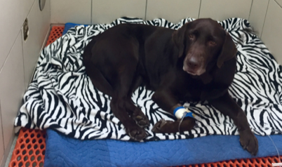
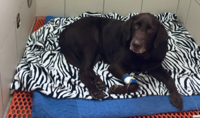
A 7 year old, female spayed, 62lb Labrador Retriever named Maple presented for ingestion of 20 gel caps of Vitamin D-3 supplement of unknown concentration- either 400IU or 5,000IU per cap. Maple had vomited three times at home, with some of the caps contained within the vomitus, and the owners called Pet Poison Helpline.
Presentation
On physical exam, Maple appeared healthy, her abdomen was not painful on palpation, and her heart rate was within normal limits. She was not lethargic or depressed at that time.
The Question
Did Maple consume enough of the supplements to cause clinical signs? Symptoms of Vitamin D toxicosis can be seen with doses as low as 0.1mg/kg, and a lethal dose in a mature dog can be as low as 2mg/kg (4). Maple weighed 28kg on entry. 1IU = 0.025mcg, and 1mcg = 0.001mg (3).
If the gel caps were 400IU: 400IU x 0.025 = 10mcg x 0.001 = 0.01mg. If she ate 20 gel caps, the total dose she consumed would be 0.2mg. For a 28kg dog, that dose would be non-toxic at 0.007mg/kg.
If the gel caps were 5,000IU: 5,000IU x 0.025 = 125mcg x 0.001 = 0.125mg. If she ate 20 gel caps, the total dose she consumed would be 2.5mg. For a 28kg dog, that dose would be non-toxic at 0.08mg/kg.
However, with the impossibility of knowing exactly what Maple consumed, the safest choice was to hospitalize her with presumed Vitamin D toxicosis.
Treatment Plan
- Hospitalize for 72 hours with IV saline to diurese the kidneys.
- UAA gel with sorbitol.
- CBC, blood chemistry panel, iStat to check free calcium, and urinalysis.
- Recheck iStat free calcium and phosphorus every 12 hours.
- Recheck CBC/chem every 24 hours to monitor renal values.
- Oral medications: furosemide, to promote calcium excretion by the kidneys (2), and famotidine, as a gastrointestinal protectant.
- Optional: adding oral prednisone (a corticosteroid), to reduce bone reabsorption of calcium and decrease absorption of calcium in the GI tract (3).
Outcome
Maple was hospitalized on IV fluids for 72 hours. Her total calcium and phosphorus levels remained within normal range for the duration of her treatment, and her renal values (BUN and creatinine) were within normal limits on exit. She was discharged with the recommendation to recheck renal values after an additional 48 hours, to ensure there would be no lasting effects from the incident.
Overview
Vitamin D toxicosis is a potentially life-threatening condition that causes increased reabsorption of calcium leading to renal damage. Clinical signs include abdominal pain, renal pain on palpation, depression, bradycardia (increased heart rate), vomiting and diarrhea (1). Once blood work is performed, findings include hypercalcemia, hyperphosphatemia, hypokalemia, and azotemia.The most common cause of vitamin D toxicity in dogs is ingestion of cholecalciferol rodenticides (4).
References:
- Tilley, Lawrence P., Francis W. K. Smith, and Lawrence P. Tilley. “Vitamin D Toxicity.” Blackwell’s Five-minute Veterinary Consult: Canine and Feline. Ames, IA: Blackwell, 2007. 1317-319. Print.
- Plumb, Donald C. “Furosemide.” Plumb’s Veterinary Drug Handbook. Stockholm, WI: PharmaVet, 2011. 454-57. Print.
- “Cholecalciferol: Rodenticide Poisoning.” Veterinary Manual. Mereck, Oct. 2014. Web. 02 Aug. 2016.
- Peterson, Michael E., and Patricia A. Talcott. “Chapter 38: Cholecalciferol.” Small Animal Toxicology. St. Louis, MO: Saunders/Elsevier, 2006. 489-98. Print.


